Revealing the Configuration of Thicker Magnets with Coherent X-rays
A soft X-ray magnetic imaging technique makes possible the study of a wide range of magnetic materials.

A solid understanding of a magnet’s nanoscale features has been critical to developing magnetic materials for applications in clean energy, sensing, computing devices, and many other technologies. But while combinations of X-ray and electron microscopy techniques can create high-resolution images of thin films of most magnetic systems, they cannot probe thicker samples of any but a few. Now, an international team led by Claire Donnelly of the Max Planck Institute for Chemical Physics of Solids have overcome this limitation with a composition-agnostic soft X-ray imaging technique for micrometer-thick magnets. The technique reveals previously inaccessible 3D textures in chiral magnets for spintronics, and naturally occurring magnetite for rock magnetism, with representing an exciting development for the study of a wider range of magnetic materials such as transition metal-based permanent magnets for energy harvesting.
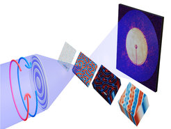
The researchers started by placing a 100-nm-thick multi-layer CoPt magnet in the soft X-ray ptychography beamline I08 at the Diamond Light Source, the UK’s national synchrotron facility. Coherent, X-rays were focused through an aperture onto the sample as it was scanned, and diffraction patterns were collected in the far-field for several overlapping positions. From these patterns, an algorithm mapped how X-rays were scattered from and absorbed by structures inside the sample. Performing these scans with different circular polarizations of the x-rays gave a similar set of mappings but with reverse contrast. The difference between mappings ultimately provided an image of the sample’s magnetic configuration via what’s known as the X-ray magnetic circular dichroism. Now, traditional techniques that measure the absorption of X-rays have probed this magnetic dichroism in absorption. Here, by measuring coherent diffraction, we also gained access to the so-called phase dichroism, described Jeffrey Neethirajan, PhD student at the Max Planck Institute for Chemical Physics of Solids and first author of the study. It turns out that in contrast to the absorption signal, which typically occurs for X-ray energies where the sample is highly absorbing, the phase signal exists for a much wider range of X-ray energies, making possible the imaging of magnetic structures in samples that would not be possible with conventional techniques.
The researchers demonstrated the technique for magnetic samples up to 1.7 μm thick and say that the capability opens the door to imaging systems that until now have not been accessible. For example, there is growing interest in topologically non-trivial three dimensional magnetic textures, that have recently been observed in chiral magnets. However, until now, it has only been possible to study such textures in thin systems, where the state is highly confined: here, the researchers were able to demonstrate that thick slabs of chiral magnetic material host unconfined states, providing an exciting outlook for the study of knot-like magnetic textures.
The internationality of the team was crucial in identifying – and demonstrating! – the impact of the technique, says Donnelly. Indeed, the team of researchers from the Max Planck Institute for Chemical Physics of Solids and the Helmholtz Centre for Materials and Energy in Germany, the Universities of Cambridge and Warwick, and the Diamond Light Source in the UK, the Paul Scherrer Institute and ETH Zurich in Switzerland, SIRUS synchrotron in Brazil, the University of Hiroshima in Japan, and Peking University in China combines expertise in synchrotron X-rays, chiral magnetism and paleomagnetism. When we spoke to our colleagues Sergio Valencia and Richard Harrison, experts on X-ray microscopy and paleomagnetism, the impact of the technique to the rock magnetism community became clear: pre-edge phase imaging provides the ability to image giant magnetofossils that until now it has not been possible to study non-destructively.
These demonstrations for chiral magnetism and rock magnetism highlight the promise of the technique, which represents an exciting advance in magnetic nanomicroscopy capabilities. And with coherence a key topic for the next generation of synchrotron radiation, as well as XFEL and lab-based sources, this use of phase dichroism highlights the importance of coherence for magnetic studies.


